£57.58 GBP inc. VAT
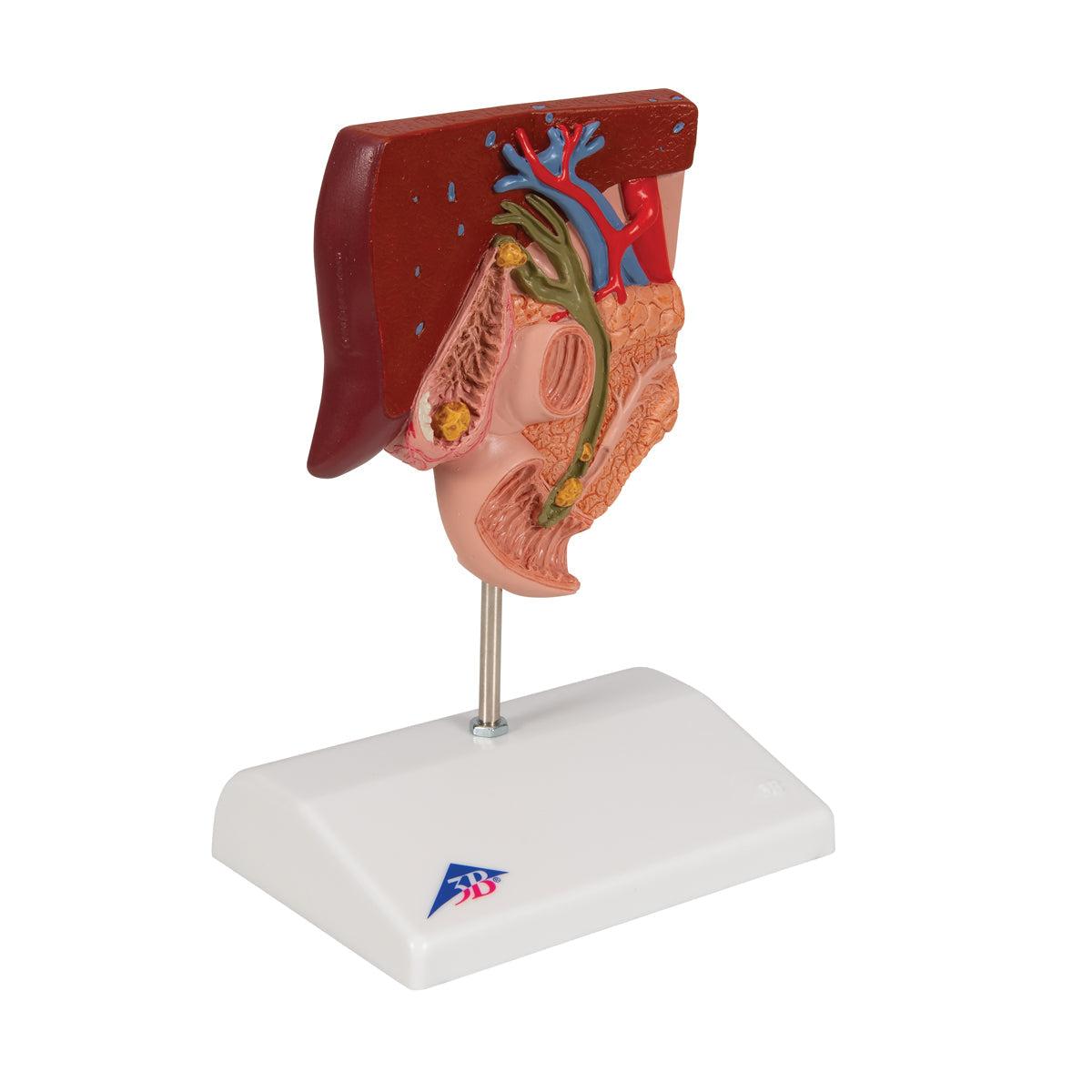
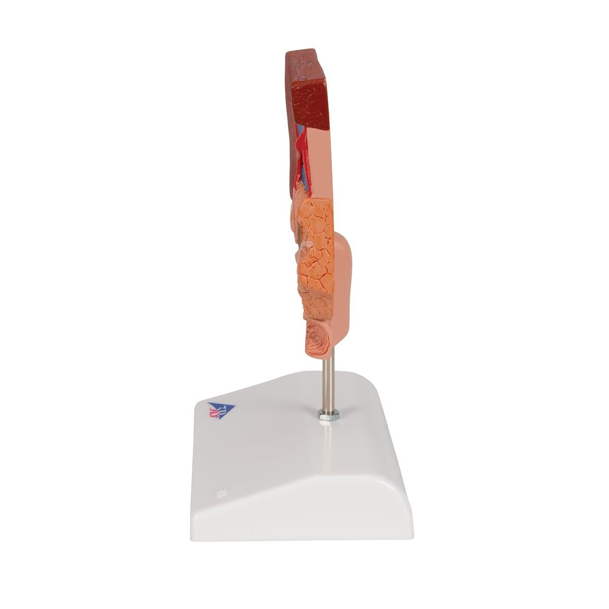
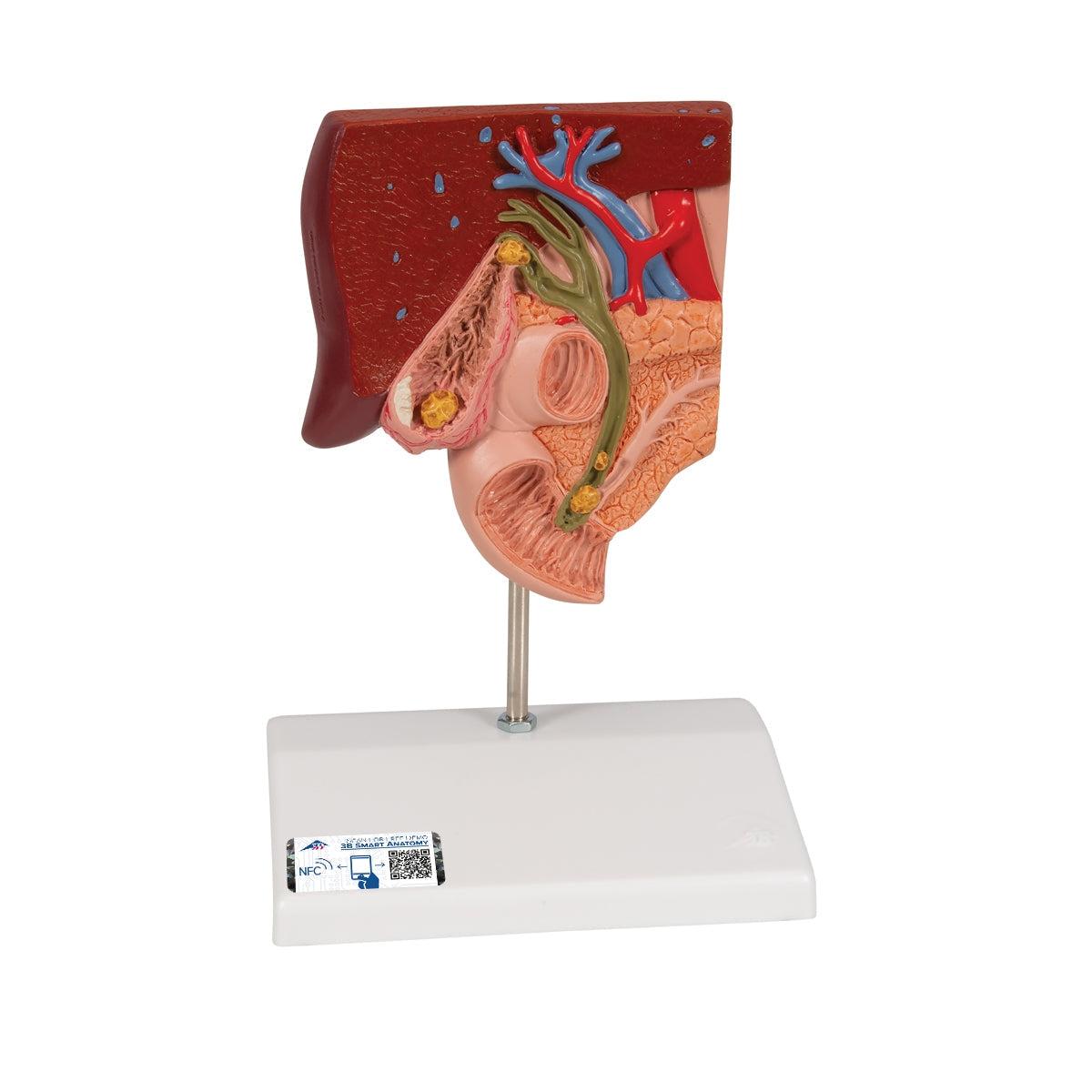
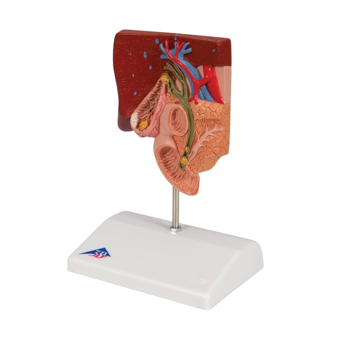
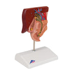
This educational gallstone model illustrates the structure of the biliary system and its adjacent areas at half the natural size. It highlights both acute cholecystitis and the tissue alterations resulting from chronic inflammation within the gallbladder wall. Gallstones are typically located in the following areas:
- In the fundus region of the gallbladder
- In the spiral valve region
- In the common bile duct area
- At the papillary opening to the small intestine
The gallstone model is securely mounted on a base.
Free Engraving Available!
Personalize Your Littmann Stethoscope with Free Engraving! Make It Uniquely Yours Today.
Use code 'FREE' at checkout.

