


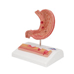
Human Stomach Section Model with Ulcers
£58.78 GBP inc. VAT
Couldn't load pickup availability
Delivery Information
Delivery Information
For orders exceeding £100 (ex VAT) within the UK mainland*, delivery is complimentary.
A nominal fee of £9.99 (ex VAT) applies to all other UK orders. Our preferred carriers include DPD and UPS, along with Royal Mail for smaller items, depending on size, value, and delivery location.
Returns & Refunds
Returns & Refunds
30-Day Hassle-Free Returns for Your Peace of Mind
Our returns policy grants you 30 days from the receipt of your item to notify our Customer Care Team if a return is necessary. Ensure the item is unused, in its original packaging, and ready for resale.
Official NHS Orders
Official NHS Orders
Ready to place an order or have inquiries about our products and services? Contact us today for exclusive NHS prices and discounts, and let’s embark on this journey towards a healthier tomorrow.
£58.78 GBP inc. VAT
This 3B Scientific® stomach pathology model illustrates different stages of gastritis, ranging from mild gastric ulcers to severe perforations. The half life-size stomach section, complete with esophagus and duodenum attachments, highlights the following stomach pathologies:
- Erythematous gastritis
- Erosive gastritis
- Hemorrhagic gastritis
- Scar formation during healing
- Atrophic gastritis
- Hypertrophic gastritis
- Bleeding ulcer
- Perforated ulcer
An additional relief model of the enlarged stomach wall features:
- Normal mucous membrane
- Acute gastritis in the antral region
- Erosive gastritis with mucosal defects
- Bleeding ulcer (eroded muscularis mucosae)
- Perforated ulcer (erosion through all stomach layers)
The stomach with ulcers is mounted on a base, ideal for educational display in classrooms or medical offices.
Free Engraving Available!
Personalize Your Littmann Stethoscope with Free Engraving! Make It Uniquely Yours Today.
Use code 'FREE' at checkout.